1/1


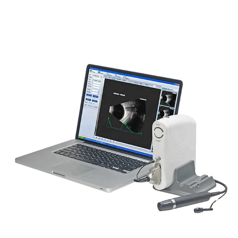
SW-2100 Ultrasound Scanner
(9 Reviews)
16 Sold
$3,800.00
1/1


Feature
1. USB interfaced ophthalmic A/B scan & image workstation is composed of main unit, A probe, B probe, image workstation and so on. The probe contacts the cornea and sends out ultrasound of 10MHz. In A scan mode, the echo wave from front surface and back surface of lens and retina is detected. The signal is processed and then we get the depth of chamber, thickness of lens and axial length. While in B scan mode, the B probe emits and receives the fan scanning waveform. After the signal procession, the transverse images of the eye are presented on the LCD. And the doctors could observe the eye disease as well as the location of the focus.
2. The ultrasound signal is pre-processed by main unit and then transported to image workstation through USB interface. The image workstation is composed of portable computer and professional ultrasound image procession software. The image process function is as powerful as to review images, measuring multiple groups of data, case output and archiving.
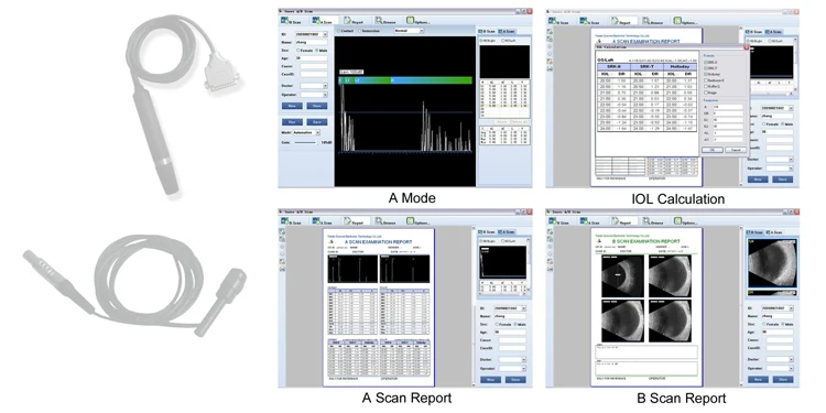
A scan:
Frequency:10MHz (imported Mini Probe) , with LEDPrecision:±0.05 mm
Measurement: Anterior chamber depth, lens thickness,vitreous body length, total length and average
Eye mode: Phakic /Aphakic /Dense / Various lOLIOL Formula: SRK-lIl, SRK-T,HOFFER-Q,
HOLLADAY,BINKHORST-II, HAIGIS
Stat. Calculation: Average and standard deviationStore: 10 Scanning results for each eye
Bscan
Frequency:10MHz,Magnetic driven, noiselessScanning Mode: Sector Scanning
Magnify: Multi continuous magnification,Real-Time magnificationResolution: Lateral ≤0.4mm; Vertical≤0.2mm
Geometry position precision: Lateral <5%; Vertical≤3%Depth:60mm
Enhance the part of vitreous body and retinaGain of probe:30dB-105dB
Scanning Angle:>=53°
Gray Scale: 256
False Color. Multi False colors. OCT False
Measurement type: multigroup distances, perimeters and areas
lmage postprocessing: multiple curves processing, Pseudo-color process-ing curve
Movies: 100 images movie review, AVIJPG format image output
Others:
Display Mode :B、B+B、B+A、AHint preset keyword
Case Search: Multi-keywords
Working Platform: Windows XP、VISTA.WINDOWs 7 etc
User-defined report template


|
A Scan
|
B Scan
|
||
|
Frequency
|
10MHz, with LED
|
Frequency
|
10MHz
Magnetic driven, noiseless
|
|
Depth
|
AL 15-35mm
|
Scanning Mode
|
Sector Scanning
|
|
Precision
|
±0.05 mm
|
Magnify
|
Multi continuous magnification,Real-Time magnification
|
|
Measurement
|
Anterior chamber depth, lens thickness
|
Resolution
|
Lateral ≤0.3mm; Vertical≤0.2mm
|
|
Geometry position precision
|
Lateral ≤10%; Vertical≤5%
|
||
|
Vitreous body length, total length and average
|
Detect depth
|
≥ 50mm
|
|
|
Gain of probe
|
20dB-105dB
|
||
|
Eye mode
|
Phakic / Aphakic / Dense / Various IOL
|
Scanning Angle
|
53°
|
|
IOL Formula
|
SRK-II, SRK-T, HOFFER-Q,HOLLADAY BINKHORST-II,HAIGIS-STD
|
Gray Scale
|
256
|
|
False Color
|
7 colors
|
||
|
Measurement type
|
Multigroup distances,
perimeters and areas
|
||
|
Stat. Calculation
|
Average and standard deviation
|
Movies
|
100 images movie review, AVI JPG.format image output.
|
|
Store
|
10 Scanning results for each eye
|
Image postprocessing
|
Multiple curves processing,Pseudo-color processing curve
|
|
Display Mode
|
B, B+B, B+A, A
|
||
|
Hint
|
Preset keyword
|
||
|
Case Search
|
Multi-keywords
|
||
|
Working Platform
|
Windows XP, VISTA, WINDOWS7
|
||
|
User-defined report template
|
With
|
||


